Week 9: Codons and Cloning
May 28, 2024
Hey blog! It’s nice to see you, welcome to the trials and tribulations of week 9– I’ve got lots to share today!
Previously, we sterilized and plated a few groups of alfalfa seeds for a germination test. Additional to those, I worked with Matias to select and plan a genetic map of our fluorescent Azospirillum colonies, which will be constructed in the following weeks. Finally, we re-fertilized all of our greenhouse groups, plus a complete survey of their germination rates and vitality.
Germination vitality assessment:
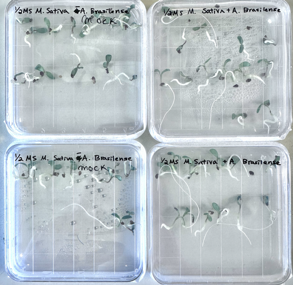
Root health and soil compatibility pose great significance to the vigor and long-term survival of plants. Because of the coarse and dry nature of MGS-1, I anticipated previously that it would be difficult to trigger germination in our alfalfa with only a natural germination schedule. Therefore, in addition to sowing seeds straight into each soil group, a supplementary trial of agar-germinated plants was conducted last week– and oh they’ve grown! (Figure 1) The control group (left) showed promising root growth, only mildly overshadowed by the Azospirillum-inoculated ones (right).

Since we are living one week post-plate, this is the time to plant them. In a group of twelve, the visually strongest seedlings of Azospirillum were transplanted into a new plot of MGS-1, mimicking the insert size and configuration of the regular experimental trial. Next to that, nine non-inoculated sprouts were transplanted into equal conditions in more MGS-1 (Figure 2).
As for our Greenhouse Check…
Considering the modifications to climate (increasing humidity, frequency of watering), the soil-germination groups planted back in Week 5 are looking unfortunately desolate. The main MGS – Azospirillum group appeared largely dried out and cakey, despite our numerous attempts to work with these harsh conditions. As such, our singular prized plant has unfortunately dried out in the fourth week, weighing an approximate 0.6 mg. This was a loss for sure, but I’m staying optimistic about my research thus far– many signs have shown that while Azospirillum did create enough nutrients for germination, long term growth requires fundamental changes to the soil’s chemical composition.
Additionally, my experimental plan is largely reliant on our trials yielding detectable amounts of plant harvests, especially an average biomass comparison across our different conditions. As such, the limited growth is posing increasing risk towards a conclusion for my paper– thus I’ve made the decision to consult alternative sources in order to mitigate the setbacks created by MGS-1’s severity.
In my literature review this week, Karthik Chinannan’s (et. al) “Effects of Mars Global Simulant (MGS-1) on Growth and Physiology of Sweet Potato: A Space Model Plant,” drew my attention to a plausible path to success. By supplementing the simulant with different concentrations of earth soil, they avoided necrosis and germination failures, and were able to analyze several components of the resulting crops’ health. The paper, published in 2024, is a promising lens for Martian agriculture; thus, I will be adapting my trials to its likeness next week in order to remediate the harsh chemical profile of these simulants.
Colonial Microscopy preparations:
On a more exciting note, practical preparation for our fluorescent bacteria (RFPB) figures has begun! Managing this process included several tasks, including:
- Germinating groups of alfalfa to be inoculated by the RFPB (below)
- Creating Electrocompetent Azospirillum colonies (details later)
- Editing MScarlet to include a start codon (details later)
- Combination of MScarlet and bacterial promoter into final plasmid (next week)
To start, we needed to create another germination plate of regular alfalfa. Contrary to the agar plates last week, these sprouts will not be transferred into soil; instead, they’ll be incubated together with our fluorescent bacteria colonies once developed, and captured via a fluorescent microscope.
This time, four tubes of selected seeds were sterilized using another Chlorine gas reaction, and the MS-media plate procedure from Week 8 was repeated for two of those tubes. The remaining two tubes were stored in a cold room– they will be plated in equal conditions next week.
Building Electrocompetent cells:
This component of the RFPB figure is largely about preparing Azospirillum to receive foreign genes (the plasmid we are developing, in this case). Here’s what I mean:
The Azospirillum we have right now are completely natural, containing their standard genome without any alterations. What we want to accomplish is the addition of foreign DNA fragments like MScarlet and a bacterial promoter, but their current state won’t allow the intake of substitutional genetic material because they are entirely stable with a natural genome.
So what researchers have done in the past is developing a cell state called “competence,” or willingness to incorporate external DNA into the cellular genetic makeup. Competence is ideal for transforming Azospirillum (and many other microbial species) because in stressful conditions, bacterial DNA will become damaged and responses will prompt the intake of floating plasmids or fragments outside of the cell membrane (Figure 3).
Our chosen method uses electroporation, the process of shocking cells with an electric pulse to produce competence. So, beginning with a culture of Azospirillum, we took periodic scans of their absorbency, the amount of light absorbed when a certain wavelength is fired through a sample of material. Absorbency has an inverse relationship with concentration, because more light absorbed through a sample means less material blocking the reception of light, and a smaller concentration.
Checking the absorbance is important because electroporation is ideal when a bacteria sample reaches the key “log-phase” of their growth state; this is when the rate of multiplication is the highest (Figure 4). Thus, waiting until their absorbance reached ~0.5 OD (Optimal Density), yielded the Azospirillum we needed– which were immediately put on ice to halt growth.
Next, cells were pelleted, meaning spun on 4,000 to 14,000 revolutions per minute in a centrifuge until collected in a clump, and suspended in a mixture of ice cold water and glycerol. These solutions were stored overnight after being frozen with liquid nitrogen (Figure 5).
On Friday, we ran the electroporation procedure, pulsing the cells in a cuvette with ~1700 Volts. Immediately after inducing competence, the Azospirillum was transferred to a liquid nutrient solution (LB – Lysogeny Broth), and refrozen.
Start Codon Addition:
And for the final RFPB update– the first stage of transformation took place this week!
Here’s some background: Our template DNA (the one we are going to add fragments to) is MScarlet, a red fluorescent protein in plasmid Matias’s stock; however, it’s currently missing an integral component of all genetic translation mechanisms, the start codon.
When proteins are synthesized from DNA, the process occurs in two steps: transcription and translation. First, an enzyme known as RNA polymerase binds to DNA, and transcribes the sequence into a complementary mRNA molecule– the form readable by ribosomes within a cell. Perceptive ribosomes are then able to compile a series of amino acids into a chain (also known as a protein) based on the information provided by mRNA, aided by a supplementary tRNA that brings miscellaneous amino acids from the cell to be turned into proteins, this is known as translation.
Any instance of translation in the cell begins with the start codon, AUG, or methionine, which signals to the ribosome where to begin synthesizing a protein.
As mentioned before, sourcing the MScarlet plasmid from Matias’s collection came with its own complications, and we need to add a start codon to it in order to produce a viable fluorescent protein code. Otherwise, the MScarlet proteins (the aspect that glows) won’t be produced from the fragment missing its start codon.
So to conclude, we ran a PCR (Polymerase Chain Reaction) to introduce methionine upstream of our MScarlet fragment. Two primers were used to amplify– primers being small fragments of DNA that bind to a template vector, hanging off by several bases that encode for additional genes. Using start-codon specific primers, we mixed the template plasmid, two primers (forward and reverse), and a Master Mix (A stock solution of many A, T, G, and C bases in suspension, as well as the enzyme Taq Polymerase). The role of Mastermix is for the Taq enzyme to use the abundance of free bases and skillfully attach them according to the primer sequence. These bases extend the template strand. (Figure)
The PCR was set to run 34 cycles in the temperatures 95 °C, 62 °C, and 72 °C at differing intervals, then stored at 12 °C. These will be tested in E. Coli cultures next week!
Whats Next?
Soon, I intend to implement the new greenhouse strategy, supplementing MGS-1 with earth soil in order to create a set of new germination plots. Further, we will introduce our new start codon MScarlet line to a culture of E. Coli, culturing them on antibiotic plates in order to verify successful transformation. If successful, these cells will be melted again and the plasmid isolated for storage!
Thanks for reading, looking forward towards Week 10!

Leave a Reply
You must be logged in to post a comment.