Final weeks: Final Results + Analysis
May 18, 2024
Welcome to my very last blog post of my senior project. These past weeks have truly gone by so fast and I’ve been able to learn and try so many new things in the lab! While there were some bumps along the way, I found that approaching problems from different angles and trying new things helped to push my project past setbacks. Now let’s dive into the final results of my project!
Last week I made a couple changes in my methodology and also incorporated the lung surfactant (CUROSURF) loading steps into my Silk Fibroin (SF) microsphere fabrication. In these past weeks I was able to carry out all 3 methods of characterization again (SEM, DLS, and LM) on new samples of microspheres to see how they reflected the changes I made in the methodology.


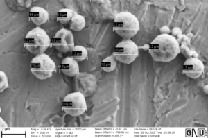
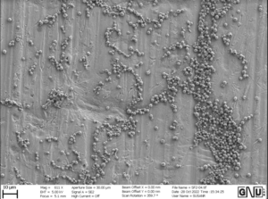
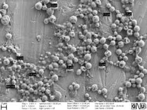
Above are the scanning electron microscopy (SEM) images from the most recent sample of SF microspheres. As you can see, there is significantly less clustering and agglomeration compared to the previous trials and the microspheres are much closer in size with most microspheres falling in the range of 1.5 to 2.7 microns. This was really great news because past research has established that to achieve targeted pulmonary drug delivery to the deep lungs, the optimal particle size for inhalation is <5 μm, and particles smaller than 3 μm have a 50–60% chance of reaching the alveolar regions. Because these SF microspheres are intended for delivering surfactant to the lung alveoli, the sizing of an average diameter 2.5 micrometers was a sufficient size. This was super exciting to see because it reflected how the changes I made in my protocol in terms of dissolution procedure and sonication settings (time and amplitude) were able to improve the final microsphere sizes and distribution.
The light microscopy images also showed a similar trend where the microspheres were more evenly spread out and similar in sizing. The dynamic light scattering (DLS) data further confirmed the beneficial changes the updated methodology made on the microspheres. The polydispersity index (PDI) of this new sample was 0.168 which was a lot closer to 0 than the initial PDI of 0.98. As I mentioned in my past blogs, a PDI closer to 0 indicates a more homogeneous sample of microspheres size, while a PDI closer to 1 indicates a more polydisperse or aggregate sample, which is something we want to avoid.
Looking at this data, I was able to successfully fabricate SF microspheres that were loaded with CUROSURF and optimize its characteristics! These findings lend insight into the potential of a more effective and efficient solution of delivering pulmonary surfactant to the lungs of newborns being affected by Neonatal Respiratory Distress Syndrome. I am super excited about the potential of my project as it not only would this method have less adverse effects on neonates, but it would also be a more efficient way to treat NRDS through the delivery of surfactant-microspheres through aerosolization.
Thanks so much for reading!

Leave a Reply
You must be logged in to post a comment.