Week 7: Data Analysis, Transformation, Validation, & Cell Culture
April 18, 2025
Last week, my mass spectrometry results finally arrived in the form of an Excel sheet with over 2000 proteins!
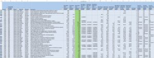
I first set some filters on the spreadsheet. I made it so that they only include proteins that are increased or decreased 2-fold while comparing between N-mut and WT; this way, I only have proteins that actually see a change between the two constructs. To do so, I entered “‘greater than or equal to 2’ or ‘less than or equal to 0.5’” in the column comparing each protein’s abundance in N-mut to WT (“NmutGFP/WTGFP”). I then created two separate sheets for when NmutGFP/WTGFP >/= 2 and </= 0.5 (ordered from highest difference to lowest difference), along with separate sheets for NmutGFP/EVGFP (empty vector GFP) and WTGFP/EVGFP.
Interestingly, my TMEM106B levels are much higher in WT than N-mut, suggesting that I didn’t add equal amounts as I should have.
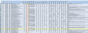
Right off the bat, Steph noticed cathepsin D, a protein involved in lysosomal protein cleavage, to be in my list. Steph then ordered a cathepsin D antibody, so I can verify whether cathepsin D really is in my samples (using my western blot membrane from last week). When I later compared my list to some pre-existing proteomics studies, the suggested association of this protein was further strengthened by Takahashi, H. et al. (2023), who also found this protein in their paper Lysosomal TMEM106B Interacts with Galactosylceramidase to Regulate Myelin Lipid Metabolism. We also found cathepsin A under the name “lysosomal protective protein” in my list.
To begin making sense of the rest of my proteins, I skimmed through and highlighted all of the proteins that had familiar or intriguing names. While highlighting, I noticed some interesting proteins: other TMEM proteins (TMED9, TMEM33, TMEM109, and of course, TMEM106B), cell cycle regulation/tumor-related proteins (CDK5, BCCIP, TPD52), proteins related to ubiquitin (NDUFAF3, UBE2S, ARIH1, USP5, UBE2D3, OTUB1), and those involved in autism, especially the mTOR pathway (LAMTOR2, CSNK2A1, FKBP1A, FMR1, ATP6V1E1, SEC13).
I then searched up lysosomal proteins and highlighted all proteins in my list that are in the lysosome. These proteins are particularly interesting because TMEM106B has been found to have cleavages in its C-terminal, which is the segment of the protein within the lysosome. A notable one that is supported by Lübke, T. (2009) includes LAMP2.
When the cathepsin D antibody arrived, I did the antibody steps on my western blot membrane again. This time, the primary antibody solution was made of 12mL blocking buffer and 4µL cathepsin D mouse antibody, and the secondary antibody solution was made of 12mL blocking buffer and 0.5µL goat anti-mouse secondary antibody. The cathepsin D antibody that we bought didn’t have a recommended western blot dilution ratio listed on its website, so I had to look through some papers’ methodologies to seek advice. However, they were not very helpful either, although I did find one paper that used a 1:3000 dilution for a different company’s cathepsin D antibody in combination with a 1:100 dilution for the one I have. 1:100 seemed too large though, so I just decided to try a 1:3000 dilution (4µL) for now, a recommended ratio for ThermoFisher’s cathepsin D antibody product.
When I checked the image results, cathepsin wasn’t visible, likely because 4µL was not enough to detect enough primary antibodies. I tried doing a 1:100 dilution this time, adding 120µL – 4µL = 116µL cathepsin D antibody to the same primary antibody solution I had used earlier. I added my newly concentrated solution to the membrane and left it overnight on the cold room shaker.
The next day, I finished the remaining antibody and wash steps and took it to the imager again. Although I couldn’t see any new signs, when I turned up the intensity very high in the red channel (which shows mouse antibodies), I could see a faint signal. When comparing it to the turned-up red channel intensity of the original membrane without cathepsin D antibody, the signals were only present in the new membrane, making it a promising sign!
Transformation
The rest of the week, I mostly spent more time on data analysis and began to work on my final project presentation, but I also began transformation to redo the entire process once again in hopes of obtaining the perfect western blot with definitive results about the cathepsin antibodies.
Cell Culture
Then on Friday, I also split some cells to prepare new cells for my next goal, to knock down the cathepsin proteins in HEK293T cells (if I find definitive cathepsin signals from the western blot) and see what happens as a result of this depletion.
Leave a Reply
You must be logged in to post a comment.