#9: Comparing ICS sources with Conventional X-Rays and Synchrotrons
May 16, 2026
In this post, I’d like to compare three major types of X-ray sources most relevant to my project: conventional laboratory/hospital X-ray tubes, synchrotrons, and Inverse Compton Scattering (ICS) sources. Although all three generate X-rays, they do so in very different ways and each has its own strengths and limitations.
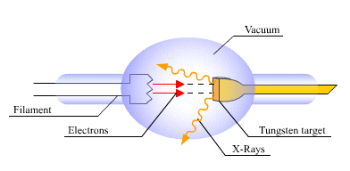
Conventional X-ray tubes are by far the most common type of X-ray source used in hospitals today. These work by accelerating electrons into a metal target, producing X-rays through a process called Bremsstrahlung radiation. These systems are very practical. They are relatively inexpensive, compact, and widely available. However, they also have important limitations. Conventional X-ray tubes produce a broad polychromatic spectrum, meaning the beam contains many different photon energies at once. Many of these photons are not useful for imaging and can contribute unnecessary radiation dose to healthy tissue. In addition, because the beam energy is spread out, these systems are not ideal for techniques such as K-edge imaging, which require very precise energy control.
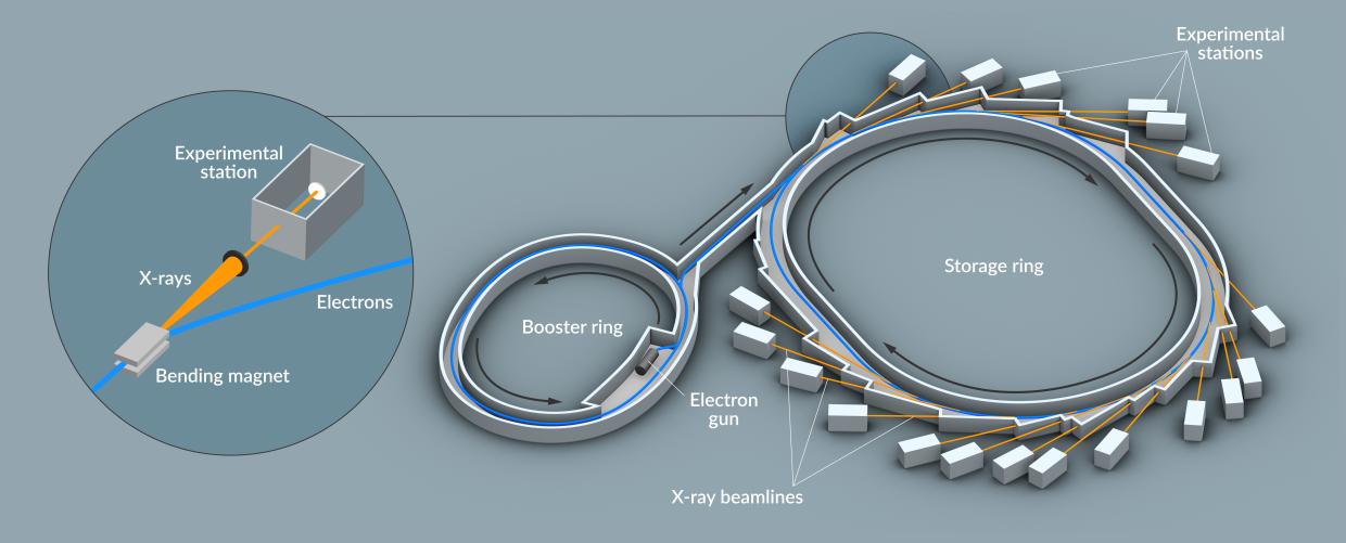
At the other extreme are synchrotrons, which are often considered the gold standard for advanced X-ray research. Synchrotrons accelerate electrons around very large circular storage rings at nearly the speed of light. As the electrons are bent by magnetic fields, they emit extremely bright and tunable X-rays. These facilities can produce very high-quality beams with excellent brightness, stability, and energy control. Because of this, synchrotrons are widely used in advanced imaging, materials science, chemistry, and biology research. However, synchrotrons also have major disadvantages. They are extremely large, expensive, and difficult to access. Many synchrotron facilities occupy entire buildings and cost upwards of hundreds of millions.
ICS sources are in between these two ends. Like synchrotrons, ICS systems can produce tunable and nearly monochromatic X-rays. However, they are much smaller and more compact. Instead of using a massive storage ring, ICS systems generate X-rays by colliding a laser beam with a high-energy electron beam. During the collision, the photons gain energy and become X-rays. One of the biggest strengths of ICS is the ability to produce narrow-energy beams that are well suited for K-edge imaging. Since the energy can be tuned very precisely, the beam can be matched to the K-edge of a contrast agent such as iodine. This improves contrast and can reduce unnecessary radiation exposure to surrounding tissue.

Leave a Reply
You must be logged in to post a comment.